Plant & Food Research Rangahau Ahumāra Kai is a New Zealand Government-owned research institute, on a mission to improve the way food is grown, harvested, prepared, and shared. Electron microscopy is a pivotal technology leveraged by the Microscopy and Imaging team in their research. The existing electron microscopy capabilities have been improved with the installation of a TESCAN CLARA Scanning Electron Microscope (SEM) with STEM (Scanning Transmission Electron Microscope) and EDS (Energy Dispersive X-ray Spectroscopy) detectors in their Microscopy Facility in Auckland. This new instrument has consolidated all their electron microscopy requirements into a single instrument as well as opening new possibilities.

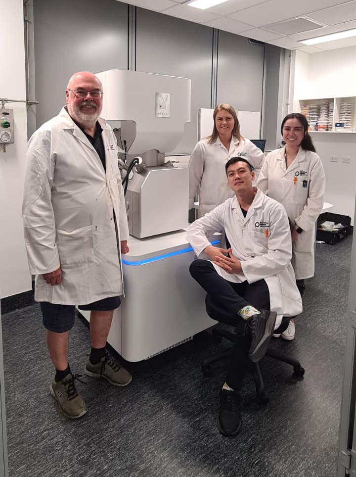
Left to right: Ian Hallett, Ria Rebstock, Andrew Chan, and Nicola Shaw.
The TESCAN CLARA is a field-emission ultra high-resolution SEM that is designed to image conductive, non-conductive, and magnetic samples making it ideally suited to imaging and analysis of a wide variety of materials. Their system specifically came equipped with cryogenic and variable pressure imaging capabilities along with EDS for elemental analysis and mapping.
When asked about the new instrument Ria Rebstock, Microscopy and Imaging Team Leader said, “when we decided to replace our aging SEM and TEM, the TESCAN CLARA with STEM detector ticked all the boxes. The increase in SEM resolution allows us to easily observe structures and answer questions we couldn’t previously. Using TESCAN’s BrightBeamTM column and beam deceleration technology enables us to image biological samples that would otherwise need coating to avoid charging, allowing us to accurately measure microscopic features such as nanocracks in starch. The addition of EDS has added new capability, which we have already leveraged for our own research, commercial clients, and industry partners. The EDS software is seamlessly integrated in the TESCAN user interface and caters to beginners and experts alike”.
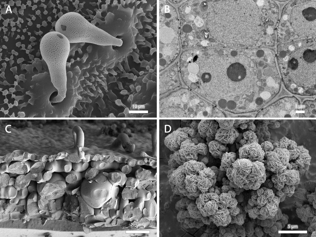
Andrew Chan, research scientist and primary instrument operator went on to say, “the instrument is so easy to use, and I have been able to customise TESCAN’s Essence user interface streamlining our workflow. New users can easily be trained to operate the CLARA in less than an hour, even people with no prior SEM experience.” Andrew added, “we have already had success imaging a diverse range of specimens as part of our research, including plant material such as fruit skins, stems, leaves, microorganisms, such as fungi, bacteria and viruses, insects, and fish. We’ve also improved the reliability of our workflows for producing TEM-like images using the retractable four quadrant BSE (backscattered electron) detector. While the sample preparation process remains largely the same, we now use robust silicon wafers instead of delicate copper or nickel grids to mount sections. We then detect backscattered electrons, which provides a TEM-like image when the contrast is inverted. Using this technique, we no longer contend with grid bars obscuring parts of our sample!”

Ria also added, “we have been very happy with the support received from both TESCAN and AXT. Everyone has been extremely accommodating and working with them has been a fantastic experience.”
AXT is TESCAN’s distributor in New Zealand and Managing Director Richard Trett commented, “to date, most TESCAN CLARA’s are being used to characterise materials. It is exciting to see how truly versatile this SEM is, and how Plant & Food Research have used it so successfully to image biological specimens. It is equally as exciting to see how they have been able to harness the improved imaging capabilities of the system so quickly and we look forward to seeing their applied research being published in the near future”.
AXT has a broad portfolio of imaging and analysis products for both industry and academia including electron microscopes, light microscopes, X-ray and molecular imaging, and a wide range of complimentary products. For more information, please visit www.axt.com.au.
