UVEX-P Automated Imager
Crystal Clear Images

Programmed scanning imager macromolecule (protein, DNA and RNA) crystallography
-
Key Features
- Automatic Scanning image entire plate or a section of a plate or select wells/drops automatically using a variety of scan profiles and light settings
- Autofocus plates can be scanned either with autofocus or without. Z value for each drop obtained by autofocus is stored for subsequent scans
- Drop Centring determine if droplets are inaccurately dispensed or migrated; quickly override if centre is off
- Extended Depth Imaging (EDI) generate a composite image from set number of Z slices and increments
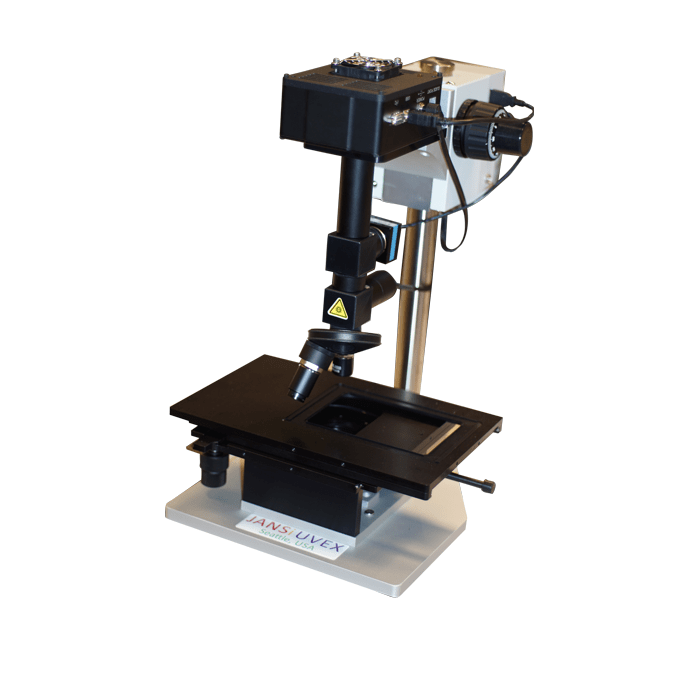
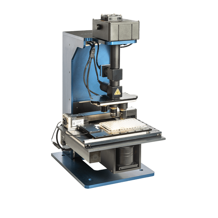
The JANSi UVEX-P offers superior specifications with motorized XYZ and objectives slider for automatic scanning of SBS and Linbro plates; includes 6 MB monochrome cooled camera, LED light sources, proprietary design for identical optical paths for brightfield and UV fluorescence images, 5x, 10x, and 20x objectives in a compact footprint.
The software for the system is a simple, intuitive interface for automatic scanning (and manual scanning of select wells if desired), with user controllable light and exposure settings, fully customised scan profiles, autofocus, drop centring, Z slicing, browser-based image management software for tracking experiments.
-
CrystalDetect Instrument Control Software
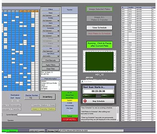
- Powerful user interface to control all aspects of the UVEX systems (UVEX-M, UVEX-P, plate hotels) and updated regularly
- Intuitive user interface is built up of layers; new layers are turned on when transiting between UVEX-M to UVEX-P and from UVEX-P to Plate Manager
-
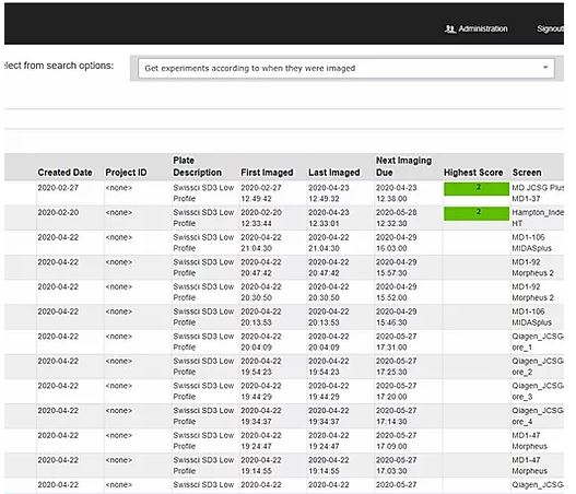
WebView Browser-based image management software
- Intuitive, multiseated and unrestricted crystal viewing and cataloguing program accessed via the user’s internet browser
- Powerful search function allows boolean searching by many different variables; screen condition, name, score, annotation, etc.
- Easy hand scoring function is available
- Entire plate can be viewed with toggling between different subwells, imaging modes and lenses
- Screen condition and plate level annotation are displayed. Comments can be added or edited
- Subwells can be inspected in greater detail, and magnification; brightfield and UV images can be overlapped
- Drop history and individual slices can be viewed; drops can be scored, and searched, well-level comments can be added
- All
- DNA/RNA
- In situ
- Micro XRD
- Molecular Biology
- Powder
- Protein
- Protein Crystallography
- SAXS/WAXS
- Small Molecule
- Thin Film
- XRD & Diffraction

