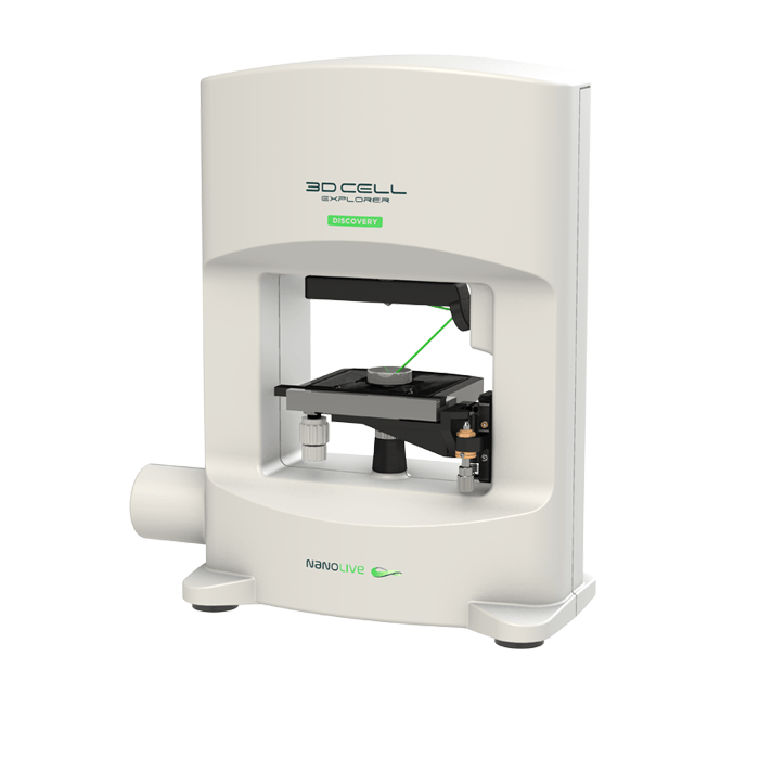
3D Cell Explorer - Live Cell Imaging Platform
Biological MicroscopyDigital MicroscopyLight MicroscopyLive Cell ImagingQuantitative Phase ImagingSuper Resolution MicroscopyTomographic Microscopy

MACROPHAGES: are present in almost all tissues. They are contributing to various processes in the healthy organism, such as development, wound healing, infection and tissue homeostasis. They can rapidly change their phenotype in response to variations in their environment. Macrophages are known for their classical function as antimicrobial phagocytes but support immune function as well by the presentation of antigens. Their research applications are vast, and in vitro assays are increasingly used in a wide range of research areas, including immunology, bacteriology and parasitology, as well as in biomedical and transplantation studies. Two advantages of macrophages in cell culture are that they are relatively easy to generate and to cultivate. find some examples in the link below
Marker-free Imaging of Cryopreserved Human M1 Macrophages
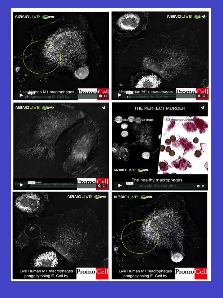
In the videos below, we can observe macrophages and T-cells interacting. Naive T-cells are being presented with antigens by the macrophages which “instruct” T-cells on what type of cells to target (such as cancer cells) and kill. During this interaction, T-cells can play a role in immune system homeostasis (Andersen, 2018) by killing the macrophage presenting the antigen. It was documented that the event is triggered by the presence of specific markers on the macrophage surface (called TRAIL and TWEAK (Kaplan et al., 2018)) telling T-cells to induce apoptosis of their fellow macrophage. The dead macrophage is then seen to be recycled by other macrophages, making space for new macrophages to be produced while keeping the same overall macrophage population.
In these videos – obtained with Nanolive’s 3D Cell Explorer – we present cryopreserved human M1 macrophages from PromoCell in cell culture (video 1). The 3D Cell Explorer allows to image these living macrophages in a novel, marker-free fashion. A special note goes to the visualization of membrane ruffling as waves arising at the leading edge of lamellipodia that move centripetally toward the main cell body. Macrophages were imaged for over 24h at a frequency of 1 image every 10 seconds. Macrophages were imaged for over 24h at a frequency of 1 image every 10 seconds.
To see more Nanolive videos related to this specific subject, please follow the link below
