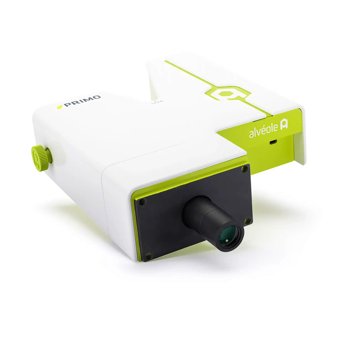
Alvéole PRIMO
Biological MicroscopyElectron MicroscopyProteinTEM

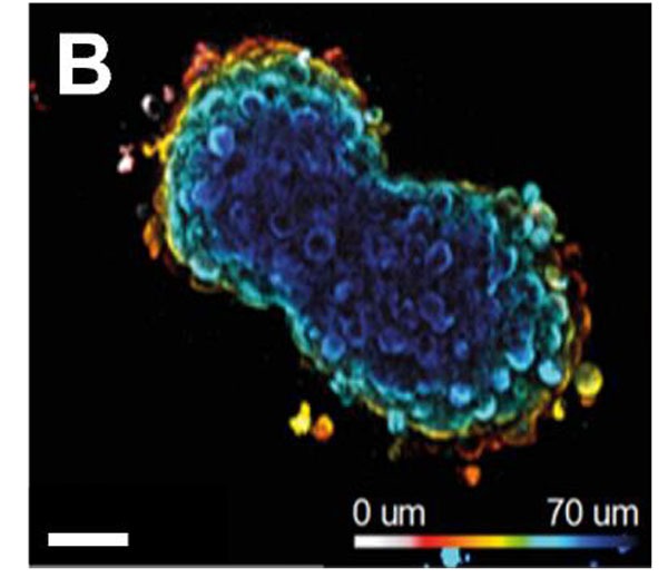
With the increasing use of spheroids in cancer research and drug screening, different techniques have been developed to produce these three-dimensional (3D) tumor cell aggregates. However, besides the fact that they are relatively labor-intensive and have limited throughput, they often lack reproducibility in the shape and size of the spheroids they form. In this application note, we show two ways of using the PRIMO contactless photopatterning system to make hundreds of very reproducible 3D cell aggregates. A first method consists in growing cells in microwells made of a non-adherent hydrogel, while the other allows their growth in 3D from a 2D micropattern.
Another limitation of current methods is the difficulty to monitor spheroid evolution or to image them because of their difficult handling. We also show by making a well-organised 3D tumor invasion assay that our techniques allow a perfect organisation of experiments in space, thus allowing easier subsequent imaging and data analysis.
This application note shows that PRIMO can be a powerful spheroid formation tool to produce reproducible 3D cell aggregates, control their shape, and organise them in space for a precise automated data analysis.
