3-D Optical Imaging for Preclinical Applications
In vivo optical imaging of preclinical mouse models is an established method that provides specific molecular information. Typical commercial optical imaging systems acquire planar, 2D images of mice that utilise either fluorescence or bioluminescence for optical contrast. Used in combination with appropriate animal models of disease, such as subcutaneous tumor models, optical imaging is used as a quick and easy “screening” approach. In concert with survival curves, intra-study sacrifice and histology, optical imaging can be an important tool in investigating disease progression and response to novel therapeutics. However, optical imaging has traditionally struggled to image deeper into the mouse, limiting its usefulness.
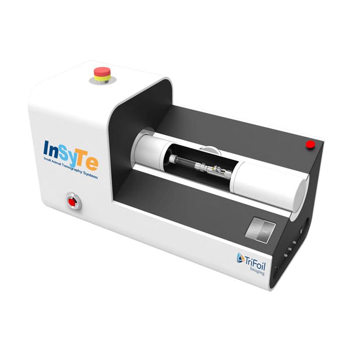
Using new architectures for imaging systems in concert with NIR fluorescent probes, we will see how imaging of targets deep within the mouse is now possible – in full 3-D and with resolution that is close to that delivered by conventional radio-nuclear modalities. We will outline some of the novel avenues of research that have been explored by teams using these advances – including oncology, inflammation, virology, cardiology, neurology, bio-distribution and probe development itself.
In vivo Optical Imaging Solutions

Fluoresence Emmision Computed Tomography
