On-demand webinar
Webinar Outline
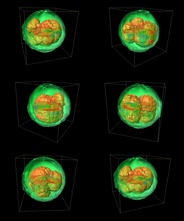
The increasing use of organoids, spheroids and bioprinted structures in translational research has necessitated a shift to unbiased objective quantitative data to better understand the biological evolution of these structures. As such researchers are needing techniques capable of imaging of cell and tissue cultures with single cell accuracy while yielding quantitative structural and dynamic information simultaneously at spatiotemporal scales, from nanometres to centimetres and milliseconds to days.
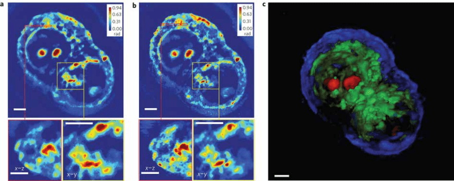
In this webinar we will describe and demonstrate two techniques; Spatial Light Interference Microscopy (SLIM) and Gradient Light Interference Microscopy (GLIM) both which provide faster and more sensitive imaging of live cells and tissues than other current technologies. Developed by Phi Optics and available as affordable add-ons to existing inverted light microscope platforms, these systems providing automated quantitative phase imaging capabilities. In this webinar you will learn how these techniques facilitate:
Watch the webinar

1) Non-invasive stable – Allows long-term investigations without cellular damage
2) Provides simultaneous information from a large field of view of many cells
3) Allows submicron levels of resolution
4) Permits 3D imaging of optically thick specimens
The capabilities of both techniques will be showcased by way of case studies. Live demonstrations can be arranged. Please contact AXT directly.


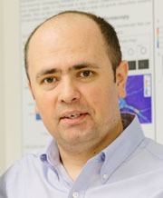
Presenter – Dr. Catalin Chiritescu
CEO and Co-Founder, Phi Optics (USA)
Dr. Chiritescu has over 20 years of experience in development of optical instrumentation, and 6 years of experience in optical metrology and ultrafast spectroscopy. He has 14 peer-reviewed publications and 5 conference presentations. He holds a PhD in Materials Science from UIUC, a MSc in Mechanical Engineering from University of Rochester, and a MSc in Physics from University of Bucharest, Romania.
