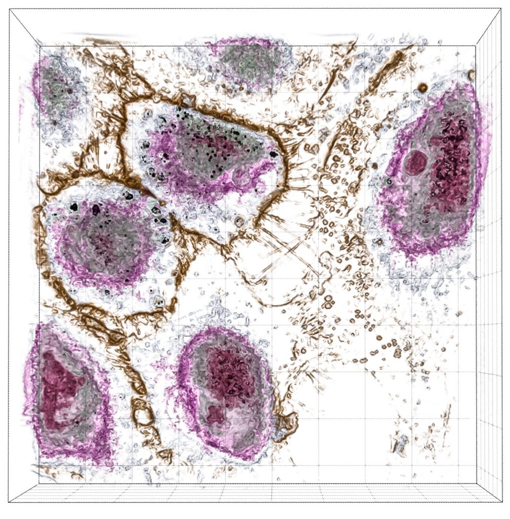
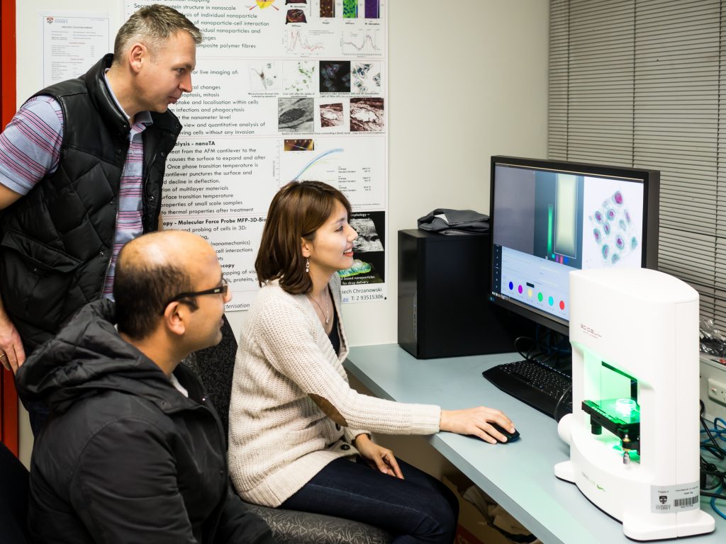
The Nanolive 3D Cell Explorer is a new and unique instrument using revolutionary technology that affords researchers a view inside living cells like never before. Australian researchers have embraced holographic tomography technology with four systems already installed and more set to follow.

Dr. Wojtek Chrzanowski from the Faculty of Pharmacy and the Australian Institute for Nanoscale Science and Technology at the University of Sydney was amongst the first adopters. His team is currently conducting research on nanotoxicity, stem cell and exosome-based therapies and cell biophysics. He first heard about the system when he attended a presentation and was hooked on the possibilities and potential from that moment and knew that the Nanolive 3D Cell Explorer was an instrument that his lab could not do without.
When asked about his initial thoughts on the system, Dr. Chrzanowski said, “I was impressed by the speed at which it could screen live cells and the ability to visualise them in 3D. For our work on nanotoxicity, we know the refractive index of the nanoparticles is different to the cells, so it should be easy to recognise and see them travelling through the cell. The technology is absolutely brilliant and we just had to have it.”
“I was impressed by the speed at which it could screen live cells and the ability to visualise them in 3D. For our work on nanotoxicity, we know the refractive index of the nanoparticles is different to the cells, so it should be easy to recognise and see them travelling through the cell. The technology is absolutely brilliant and we just had to have it.”
Dr. Wojtek Chrzanowski
Their system was installed earlier this year and operators are able to generate images within 45 to 60 minutes of training thanks to the simplicity of its design and the “super-easy” user-friendly interface. When asked about results they had produced thus far, he replied, “our work on nanoparticle uptake is progressing very well and we have been able to produce top notch images that support our hypotheses. Based on these results we will be submitting a manuscript very soon. One of my students will also be presenting this data at the International Particle Toxicity Conference in Singapore later this year. We are also looking at how treatments affect tissue fibrosis at the cellular level using the 3D Cell Explorer and results achieved so far have been very positive.”
“In the near future we will be looking to carry out nanotoxicity studies in an incubator with the aim of visualising cellular interactions in real time to see the uptake of nanoparticles and their transport path, as well as any changes in cellular morphology.”

Another advantage of using the 3D Cell Explorer is the ability to digitally stain samples using the STEVE software interface. Dr. Chrzanowski commented, “digital staining is a definite advantage as it provides cost and time savings. It will become more of an advantage in the future as we generate more data on refractive indices which will allow us to better utilise STEVE.”
Currently, their system has 6 core users with numerous additional users from outside their group of which some are looking at original and unique research projects. Dr. Chrzanowski said of his experience with the 3D Cell Explorer so far, “it is absolutely fantastic! Its ease of operation, intuitive nature, compact size, rapid imaging and no need for stains make this a system I would certainly recommend and it would certainly support many different kinds of research. In fact, this system would be valuable for secondary students studying biology too.”
