On-Demand Webinar
Overview
Intravital microscopy is an invaluable imaging technique to visualise various in vivo cellular-level dynamics in a live animal. Intravital imaging in a natural physiological microenvironment can provide valuable insights in the dynamic pathophysiology of human diseases compared with conventional histological observation of ex vivo sample or in vitro culture systems. Over the last decade, intravital microscopy has become an indispensable technique and has enabled in vivo visualisation of numerous biological processes. In doing so, it has also facilitated the development of new therapeutics and diagnostics.
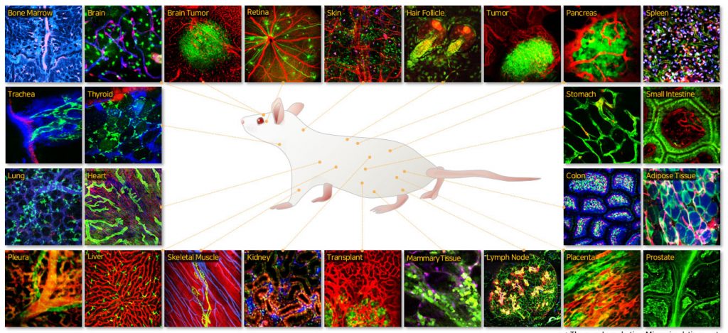
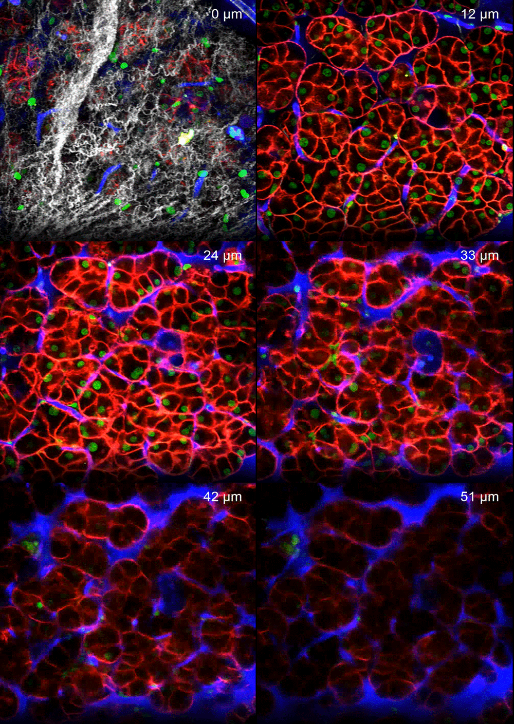
In this talk, IVIM Technology’s all-in-one real-time intravital two-photon and confocal microscopy system will be introduced. Optimised for in vivo cellular-level imaging it can acquire real-time multi-color sub-micron-resolution images in the internal organs of live animal models with automatic motion compensation functionality. Intravital imaging of various organs including skin, liver, spleen, pancreas, kidney, small intestine, colon, retina, lung, heart, lymph node, bone marrow, and brain will be shown. Recent studies utilising this imaging technique to investigate dynamic cellular-level pathophysiology of various human diseases are presented. A longitudinal intravital imaging study of cerebral microinfarction is described.

Presenter – Prof. Pilhan Kim

Dr. Kim is a Professor at Korea Advanced Institute of Science and Technology (KAIST) in the Graduate School of Medical Science and Engineering and authored over 100 publications as well as 16 patents. He is also the CEO/CTO at IVIM Technology and holds a BC and PhD in electrical engineering. He has also served as a Research Fellow at Harvard Medical School and Massachusetts General Hospital in the USA.


